Saturday, 28 December 2013

Questions about Pacemakers - Best Cardiac Hospitals in India

Heart rhythm problems (arrhythmia) occur when the electrical impulses produced by your heart that coordinate heartbeat do not function properly, causing your heart to beat too quickly, too slowly, or irregularly.Age increases the probability of experiencing an arrhythmia. It can occur in people who do not have heart disease.Some heart arrhythmias are harmless, though some types, such as ventricular tachycardia (fast heart rates), are serious and even life threatening.
Pacemakers represent one of the earliest and most successful nonpharmacological therapy for arrhythmias. Millions of pacemakers have been implanted since the very first pacemaker was implanted. Drugs are no longer used except in the very acute setting before implantation of a temporary or permanent pacemaker.

A cardiac pacemaker is a device that is used to regulate the heart rate.

If you have been found to have a heartbeat that is too slow, a pacemaker can be implanted in the body to take over the function. This small electronic device automatically monitors and regulates the heartbeat, by transmitting electrical impulses to stimulate the heart when it is beating too slowly. A pacemaker consists of a pacing lead and a pulse generator. Single chamber pacemakers have only a single lead while dual chamber pacemakers have two leads with one lead in the atrium and the other in the ventricle. Dual chamber pacemakers are more physiological but more expensive.

The indications of pacing are now well established. The most important indication of pacing however remains complete heart block and the sick sinus syndrome which account for 95% of the indication for pacemakers implanted in Singapore. During the last pacemaker survey in 2005 in Singapore, the implant rate was 91 per million. With our ageing population, we can expect that the need for pacemaker implantation in Singapore will rapidly increase.


Single chamber pacemakers are pacing systems that use one lead in either the right atrium or the right ventricle of your heart.
A single lead in the right atrium is commonly used in conditions where the normal pacemaker of the heart is not working adequately, such as in the case of sick sinus syndrome. Atrial pacing is used when the sinus node is sending out signals that are too slow or irregular. However, to use this method of pacing, the rest of the heart's normal conduction system must be functioning normally.
More commonly, the single lead is placed in the right ventricle to help correct a slow or irregular heart beat. This is most often the case when the electrical flow is slowed or blocked in the region of the atrio-ventricular (A-V) node and the normal impulses from the atria cannot reach the ventricle. This would result in too slow a heart beat. The pacemaker system would keep the heart beating at a steady rate

Dual chamber pacemakers are pacemaker systems that use a lead in the right atrium as well as the right ventricle (figure 6). This type of pacing most closely mimics the heart's normal conduction pattern by pacing sequentially from atria to ventricle thus maximizing the heart's pumping ability. By having a lead in both the atria and ventricle the pulse generator is able to continuously regulate the heart's electrical activity in both chambers. These are the most commonly used pacemakers at the present time.

Commonly asked questions about pacemakers

Will I need to make any lifestyle changes after my pacemaker is implanted?
There are no significant lifestyle changes that you will need to make as a result of having a pacemaker implanted. Most patients resume their normal activities soon after implantation. Specific issues or concerns should be addressed with your pacemaker physician or nurse.
How often will I need to have my pacemaker checked?
Your pacemaker system will need to be evaluated by your pacemaker physician, nurse, or your local cardiologist's office at least twice yearly. A special computer called a programmer is used to perform a comprehensive evaluation of your pacemaker system. The programmer has a wand (like a computer mouse) that is used to communicate with the pacemaker. The wand is placed on your chest directly over the pulse generator and a radio wave signal is used to send and receive information from the pulse generator. Changes in the pacemaker settings can be done via this method as well. A complete assessment of the pacemaker's sensing and pacing functions, battery life, and diagnostic information is obtained, which enables your pacemaker physician/nurse to fine tune your care.
How is the battery changed?
The battery that is used to power your pulse generator is tightly sealed within the metal shell of the device. Therefore, when the battery's energy is depleted a whole new pulse generator must be implanted. The skin over the pulse generator site is numbed up with local anesthetic. You may also receive a light sedative through a intravenous line to help you relax. A new incision is made in the skin and the pacemaker pocket is opened. The pulse generator is removed and lead(s) disconnected. At this time the lead(s) are hooked to a special analyzer that evaluates the lead(s) for any evidence of potential malfunction. A new pulse generator is then attached to the lead(s) and the system is reimplanted in the same pocket. The incision is sutured (sewn) together and a small dressing applied. Most patients can go home the same day as their procedure.
Can I use a microwave?
Microwave ovens will not interfere with the proper functioning of your pacemaker. You can use a microwave oven without concern.
Can I use a cell phone?
It is possible that a cellular phone might interfere with the normal functioning of your pacemaker. The interaction is temporary, however, and will only affect the pacemaker during the time that your cellular phone is close to your pacemaker. To avoid this potential interference, it is recommended that you hold the cellular phone on the opposite side of your body away from the pacemaker. You should also not store your cellular phone in your breast pocket.You should always try to maintain a distance of at least 6 inches between your cellular phone and your pacemaker system.
Do I have to take any precautions at the airport?
If you were to walk through the metal detector at the airport, it will not harm you nor your pacemaker. However, because the pacemaker is encased in a metal shell, it is possible that the pacemaker may set off the security alarm. To avoid this problem, it is generally recommended that you show your pacemaker identification card to the security agent and inform him/her that you have an implanted pacemaker system. They should let you pass around the metal detector. If the airport security wants to scan you with the "hand wand", they can everywhere except over the device. This information also pertains to any metal detector such as at a courthouse or federal building.


Thursday, 26 December 2013

Heart Defects Treatment Options available in India – Types and Treatment for Holes in the Heart


What Are Holes in the Heart?
Holes in the heart are simple congenital heart defects. Congenital heart defects are problems with the heart's structure that are present at birth. These defects change the normal flow of blood through the heart.
Your heart has two sides, separated by an inner wall called the septum. With each heartbeat, the right side of your heart receives oxygen-poor blood from your body and pumps it to your lungs. The left side of your heart receives oxygen-rich blood from your lungs and pumps it to your body. The septum prevents mixing of blood between the two sides of the heart. However, some babies are born with holes in the upper or lower septum.
A hole in the septum between the heart's two upper chambers is called an atrial septal defect (ASD). A hole in the septum between the heart's two lower chambers is called a ventricular septal defect (VSD).
ASDs and VSDs allow blood to pass from the left side of the heart to the right side. This means that oxygen-rich blood can mix with oxygen-poor blood. As a result, some oxygen-rich blood is pumped to the lungs instead of out to the body.
Over the past few decades, the diagnosis and treatment of ASDs and VSDs have greatly improved. Children who have simple congenital heart defects can survive to adulthood and live normal, active, and productive lives because their heart defects close on their own or have been repaired.

Types of Holesin the Heart

Atrial Septal Defect

An atrial septal defect (ASD) is a hole in the part of the septum that separates the atria (the upper chambers of the heart). This hole allows oxygen-rich blood from the left atrium to flow into the right atrium instead of flowing into the left ventricle as it should. This means that oxygen-rich blood gets pumped back to the lungs, where it has just been, instead of going to the body.


Cross-Section of a Normal Heart and a Heart with an Atrial Septal Defect


The normal structure and blood flow in the interior of the heart. Figure B shows a heart with an atrial septal defect. The hole allows oxygen-rich blood from the left atrium to mix with oxygen-poor blood from the right atrium. An ASD can be small or large. Small ASDs allow only a little blood to flow from one atrium to the other. Small ASDs don't affect the way the heart works and don't need any special treatment. Many small ASDs close on their own as the heart grows during childhood.
Medium to large ASDs allow more blood to leak from one atrium to the other, and they're less likely to close on their own. Most children who have ASDs have no symptoms, even if they have large ASDs.
The three major types of ASDs are:
  • Secundum. This defect is in the middle of the atrial septum. It's the most common form of ASD. About 8 out of every 10 babies born with ASDs have secundum defects. At least half of all secundum ASDs close on their own. However, this is less likely if the defect is large.
  • Primum. This defect is in the lower part of the atrial septum. It often occurs along with problems in the heart valves that connect the upper and lower heart chambers. Primum defects aren't very common, and they don't close on their own.
  • Sinus venosus. This defect is in the upper part of the atrial septum, near where a large vein (the superior vena cava) brings oxygen-poor blood from the upper body to the right atrium. Sinus venosus defects are rare, and they don't close on their own.

AtrialSeptal Defect Complications

Over time, if an ASD isn't repaired, the extra blood flow to the right side of the heart and lungs may cause heart problems. Usually, most of these problems don't show up until adulthood, often around age 30 or later. Complications are rare in infants and children.
Possible complications include:
  • Right heart failure. An ASD causes the right side of the heart to work harder because it has to pump extra blood to the lungs. Over time, the heart may become tired from this extra work and not pump well.
  • Arrhythmias (ah-RITH-me-ahs). Extra blood flowing into the right atrium through an ASD can cause the atrium to stretch and enlarge. Over time, this can lead to arrhythmias (irregular heartbeats). Arrhythmia symptoms may include palpitations or a rapid heartbeat.
  • Stroke. Usually, the lungs filter out small blood clots that can form on the right side of the heart. Sometimes a blood clot can pass from the right atrium to the left atrium through an ASD and be pumped out to the body. This type of clot can travel to an artery in the brain, block blood flow, and cause a stroke.
These problems develop over many years and don't occur in children. They also are rare in adults because most ASDs either close on their own or are repaired in early childhood.

VentricularSeptal Defect

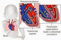
A ventricular septal defect (VSD) is a hole in the part of the septum that separates the ventricles (the lower chambers of the heart). The hole allows oxygen-rich blood to flow from the left ventricle into the right ventricle instead of flowing into the aorta and out to the body as it should.

Cross-Section of a Normal Heart and a Heart With a Ventricular Septal Defect


The normal structure and blood flow in the interior of the heart. two common locations for a ventricular septal defect. The defect allows oxygen-rich blood from the left ventricle to mix with oxygen-poor blood in the right ventricle. An infant who is born with a VSD may have a single hole or more than one hole in the wall that separates the two ventricles. The defect also may occur by itself or with other congenital heart defects.
Doctors classify VSDs based on the:
  • Size of the defect.
  • Location of the defect.
  • Number of defects.
  • Presence or absence of a ventricular septal aneurysm-a thin flap of tissue on the septum. This tissue is harmless and can help a VSD close on its own.
VSDs can be small or large. Small VSDs don't cause problems and often may close on their own. Because small VSDs allow only a small amount of blood to flow between the ventricles, they're sometimes called restrictive VSDs. Small VSDs don't cause any symptoms. Medium VSDs are less likely to close on their own. They may require surgery to close and may cause symptoms during infancy and childhood.
Large VSDs allow a large amount of blood to flow from the left ventricle to the right ventricle. They're sometimes called nonrestrictive VSDs. A large VSD is less likely to close completely on its own, but it may get smaller over time. Large VSDs often cause symptoms in infants and children, and surgery usually is needed to close them.
VSDs are found in different parts of the septum.
  • Membranous VSDs are located near the heart valves. These VSDs can close at any time.
  • Muscular VSDs are found in the lower part of the septum. They're surrounded by muscle, and most close on their own during early childhood.
  • Inlet VSDs are located close to where blood enters the ventricles. They're less common than membranous and muscular VSDs.
  • Outlet VSDs are found in the part of the ventricle where blood leaves the heart. These are the rarest type of VSD.
For more information visit:          http://www.medworldindia.com        
                    
https://www.facebook.com/medworld.india

Please scan and email your medical reports  to us at care@medworldindia.com and we shall get you a Free Medical Opinion from India’s Best Doctors.

Call Us : +91-9811058159
Mail Us : care@medworldindia.com


Saturday, 21 December 2013

Benefits of Heart Valve Repair and Heart Valve Replacement Surgery at World Best Hospitals in India

 Heart Valve repair and Heart Valve Replacement Surgery


Surgical

  • A commissurotomy is surgery to open up valves that have thickened and are perhaps stuck together. The valve is opened by cutting the points where the leaflets of the valve meet. Commissurotomy is a type of valvuloplasty or valve re-shaping. 
  • Annuloplasty is a technique to repair an enlarged annulus, a ring of fibrous tissue at the base of the heart valve. To repair the annulus, sutures are sewn around the ring to make the opening smaller. Alternatively, a ring-like device is attached around the outside of the valve opening. The ring provides support to the valve so it can close more tightly. 
  • A surgeon may reshape a valve by cutting out a section or sections of a leaflet and then sewing the leaflet back together. 
  • Decalcification is surgery to remove calcium buildup from the leaflets. 
  • Valves are supported by cords (called chordae tendineae) and papillary muscles. If these are stretched or weak, the valve may not close properly. By replacing or shortening the cords, the valve will be strengthened and able to close properly. 
  • If there are holes or tears in the leaflets of the valve, a surgeon may repair them with a tissue patch.
Unlike conventional surgery, minimally invasive surgery does not involve sawing through the breastbone and opening the chest. The surgeon watches the heart on a video screen and operates using long-handled surgical tools inserted through small incisions. In some cases, robotic arms are used. Minimally invasive valve repair is only available in some hospitals. It is also referred to as endoscopic or robotic heart surgery.


Non-surgical valve repair
Percutaneous or catheter-based procedures are done without any incisions in the chest or stopping the heart. Instead, a thin flexible tube called a catheter is inserted into a blood vessel in the groin or the arm and then threaded up into the interior of the heart.
  • Percutaneous or balloon valvuloplasty is used in people with stiffened or narrowed (stenosed) pulmonary, mitral or aortic valves (more commonly for the mitral than the aortic valve). A balloon tip on the end of the catheter is positioned in the narrowed valve and inflated to enlarge the opening or to crack open calcified tissue. 
  • Several methods of percutaneous mitral valve repair are being developed. These procedures are still in the developmental phase and are available in a limited number of centres. One example is edge-to-edge repair, which can be used in the case of a very leaky mitral valve in a patient who is considered high risk for a surgical repair or replacement. A delivery catheter holding a clip is advanced through the femoral vein from the groin into the left side of the heart, under general anesthesia. The clip is positioned beyond the leaky valve in an open position and then pulled back so that it catches the flaps of mitral valve. Once closed, the clip holds the leaflets together and stops the valve from leaking.
Valve Replacement


Replacement is more commonly used to treat aortic valves or severely damaged mitral valves. There are two kinds of valves that are used for valve replacement and you should talk with your doctor about which type is best for you.
  • Mechanical valves are made from durable metals, carbon, ceramics and plastics. A fabric sewing ring is used to attach the valve to the tissues in the patient’s heart. The major advantage is durability. However, blood thinners must be taken the rest of the patient’s life to prevent blood clots. The valve makes a soft clicking sound when it floats shut, which may bother some patients. People generally adjust quickly to this sound. 
  • Biological valves are made from:
    • Animal tissue, either an actual pig valve or a bovine pericardial engineered valve (a xenograph).
    • Human tissue of a donated heart (an allograft or homograft), used most often to replace infected valves.
    • A patient’s own tissues (an autograft). A Ross Procedure (also called a Switch Procedure) involves taking the patient’s normally functioning pulmonary valve and using it to replace a diseased aortic valve. The pulmonary valve is then replaced with a donated (homograft) pulmonary valve.
Biological valves are not as durable as mechanical valves and may need to be replaced between 5 and 15 years. Patients with biological valves will need to take blood thinners in the short term.
The most advanced technology for minimally invasive heart surgery is now available in India. Robotic da Vinci Surgical System combines superior 3D visualization along with greatly enhanced dexterity, precision and control in an intuitive, ergonomic interface with breakthrough surgical capabilities.


In Robotic Cardiac Surgery, robotic assist the heart surgeon in performing surgery through tiny incisions. The surgeon has to sit in an operating room where he controls the instruments while looking at 3D images from the camera which is inside the patient. Various procedures can be performed in this case such as mitral valve replacement or repair, correction of HOCM etc. Use of Robotic Surgery in Cardiology has benefits as it involves less pain, scarring and risk that are otherwise involved. The surgeon can perform surgery through tiny openings in the chest, cracking the breastbone and spreading the ribs.


Heart Surgery in India with the Best Heart surgeons in India is what medical tourists coming to India look for and is delivered at best cardiac hospitals in India . No other destination in the world can match the unparalleled clinical excellence, technology infrastructure and cost advantage offered by MedWorld India affiliated heart hospitals in India to deliver 99 percent and above successful outcomes for heart surgery.
 Our aim is to reduce the deadly consequences of cardiovascular diseases through innovative therapies for patient care. Our healthcare professionals at state of the art, heart specialty hospitals have a dedicated team of Cardiac surgeons and cardiologists who work in tandem to provide comprehensive, multidisciplinary care to patients suffering from heart ailments.

MedWorld India Affiliated Best Heart Hospitals in India offer: - The ultimate destination for international patients who want World Best, Affordable Heart Surgery
India offers great value proposition to patients looking for affordable Heart surgery or Cardiac surgery abroad as not only the cost of valve replacement in India is very low, the facilities and infrastructure is most modern and is counted among the best heart hospitals in the world. Cardiac surgeons now use most advanced minimally invasive heart valve replacement or repair surgery techniques for eligible patients.

For more information visit:          http://www.medworldindia.com     
                                                          
https://www.facebook.com/medworld.india

 Please scan and email your medical reports  to us at care@medworldindia.com and we shall get you a Free Medical Opinion from India’s Best Doctors.

  Call Us : +91-9811058159
  Mail Us : care@medworldindia.com

Thursday, 19 December 2013

Different Techniques used for fixing congenital Defects of the Heart in Children - Pediatric Cardiac Surgery India

Paediatric Heart Surgery in India with the Best Paediatric Heart surgeons in India is what you would want if your little one is suffering from any heart ailment. MedWorld India affiliate Paediatric heart surgeons in India are among the top paediatric heart specialists and offer best in paediatric heart care. Affordable Pediatric Heart Surgery in India at Best Cardiac Hospitals with Top Pediatric Cardiac Surgeons in India with MedWorld India.

Sometimes heart surgery in children is required for repairing defects in the heart which a child might have born with (known as congenital heart defects) and heart diseases which he gets after birth, which require surgery. Pediatric Cardiac Surgery deals with operative procedures in the newborn and unborn children and youngsters suffering from cardiac dysfunctions, structural, functional and rhythm-related issues of the heart also.

Major Types of Cardiac Defects in Children :-


Thoracotomy: This Procedure is used for some type of heart defect repairs, wherein the incision is made on the side of the chest, between the ribs. It is also called a closed-heart surgery. This surgery is done with the help of special instruments and a camera.In one kind of procedure a few small tubes are inserted into an artery in the leg and pass them up to the heart. Only some heart defects can be repaired in this fashion.

Heart Surgery in India with the Best Heart surgeons in India is what medical tourists coming to India look for and is delivered at Best Cardiac Hospitals in India . No other destination in the world can match the unparalleled clinical excellence, technology infrastructure and cost advantage offered by MedWorld India affiliated heart hospitals in India to deliver 99 percent and above successful outcomes for heart surgery.

 
Our aim is to reduce the deadly consequences of cardiovascular diseases through innovative therapies for patient care. Our healthcare professionals at state of the art, heart specialty hospitals have a dedicated team of Cardiac surgeons and cardiologists who work in tandem to provide comprehensive, multidisciplinary care to patients suffering from heart ailments.

MedWorld India Affiliated Best Heart Hospitals in India offer: - The ultimate destination for international patients who want World Best, Affordable Heart Surgery

For more information visit:          http://www.medworldindia.com       
                                                          
https://www.facebook.com/medworld.india


Please scan and email your medical reports  to us at care@medworldindia.com and we shall get you a Free Medical Opinion from India’s Best Doctors.


Call Us : +91-9811058159
Mail Us : care@medworldindia.com

Thursday, 12 December 2013

Latest and Best Robotic Heart Surgery in India

Robotic Heart Surgery are now widely used to provide patients with less pain and faster recovery time. Limitations in surgical equipment have prohibited their use during heart bypass surgery  until now.

 Da Vinci Robotic System
The advancement of computerized robotic technology is one of the most exciting breakthroughs in surgery to date, enabling surgeons to perform complete, bypass surgery less invasively. Penn Cardiac Care at the Hospital of the University of Pennsylvania is proud to be the only hospital in Philadelphia and one of only 12 in the nation invited to participate in a study to determine the effectiveness of this revolutionary procedure.


Robotic Heart Surgery in India is the latest and most advanced technique in the world to perform heart surgery with greatest precision Robot Assisted Heart Procedures are done to treat a variety of conditions:


Heart Surgery in India with the Best Heart surgeons in India is what medical tourists coming to India look for and is delivered at best cardiac hospitals in India . No other destination in the world can match the unparalleled clinical excellence, technology infrastructure and cost advantage offered by MedWorld India affiliated heart hospitals in India to deliver 99 percent and above successful outcomes for heart surgery.

Our aim is to reduce the deadly consequences of cardiovascular diseases through innovative therapies for patient care. Our healthcare professionals at state of the art, heart specialty hospitals have a dedicated team of Cardiac surgeons and cardiologists who work in tandem to provide comprehensive, multidisciplinary care to patients suffering from heart ailments.


MedWorld India Affiliated Best Heart Hospitals in India offer: - The ultimate destination for international patients who want World Best, Affordable Heart Surgery